

Presentation of CIDP can vary greatly across the patient population1
There are multiple variants of chronic inflammatory demyelinating polyneuropathy (CIDP), and it can be classified into typical CIDP and CIDP variants. Common symptoms include weakness, numbness, and pain; however, symptoms can vary greatly from patient to patient, making diagnosing typical CIDP and CIDP variants of the disease challenging.1,2
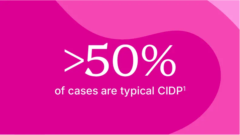
Proximal and distal symmetric weakness with sensory impairment and absent or hyporeflexic reflexes. Symptoms worsen progressively over >8 weeks and may follow a relapsing course
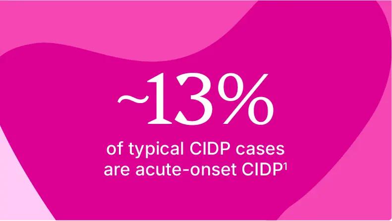
Proximal and distal symmetric weakness with sensory impairment and absent or hyporeflexic reflexes. Rapid worsening within 4 weeks, which may mimic Guillain-Barré syndrome (GBS) in early stages. However, patients with CIDP differentiate from those with GBS because they continue to show deterioration for more than 8 weeks or relapse at least 3 times. Patients with acute onset CIDP are able to walk and are less likely to have facial weakness
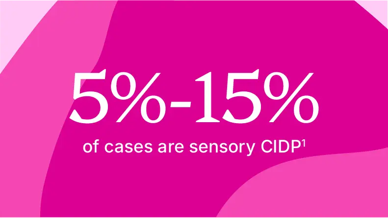
Predominantly sensory symmetric presentation with ataxia and generalized reduced sense to vibration and position changes
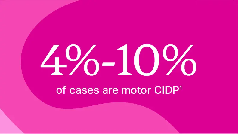
Distal and proximal symmetric motor impairment
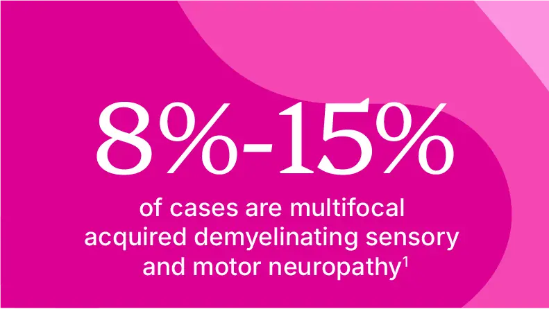
Asymmetric sensory and motor deficits, distal greater than proximal, with upper extremities more affected than lower

Distally predominant symmetric impairment, sensory greater than motor, with lower extremities more affected than upper
Understanding CIDP and CIDP variants is essential to reaching an accurate diagnosis and appropriate clinical management of the disease. Each variant presents distinct clinical features, electrophysiological patterns, and disease progression timelines that may impact patient outcomes.1
These are not the only presentations of CIDP.
It’s important to keep multiple factors in mind when diagnosing CIDP and its variants1,2
Factors that may impact diagnosis include:
Clinical variability1,2
CIDP presents along a spectrum, from typical symmetric presentations to rare variants.
Some patients, particularly those with severe axonal damage, don't fulfill standard diagnostic criteria. Diagnosing CIDP is challenging due to its heterogeneity and lack of specific diagnostic tests and validated biomarkers.
Electrodiagnostic complexity1-3
While European Academy of Neurology/Peripheral Nerve Society criteria provide standardized thresholds, interpretation can be complex. In some cases, working with a multidisciplinary team may be recommended, particularly in borderline cases or when patients don't clearly meet the criteria.
Laboratory limitations1,3,4
No validated biomarker tests for CIDP and its variants currently exist. No universal cerebrospinal fluid protein upper reference limit exists. Supportive tests help but cannot replace clinical and electrodiagnostic criteria.

References: 1. Roman-Guzman RM, Martinez-Mayorga AP, Guzman-Martinez LD, Rodriguez-Leyva I. Cureus. 2025;17(1):e76749. doi:10.7759/cureus.76749 2. Broers MC, Bunschoten C, Drenthen J, et al. Eur J Neurol. 2021;28:2065-2073. doi:10.1111/ene.14796 3. Stino AM, Naddaf E, Dyck PJ, Dyck PJB. Muscle Nerve. 2021;63(2):157-169. doi:10.1002/mus.27046 4. Allen JA. Neurol Ther. 2020;9(1):43-54. doi:10.1007/s40120-020-00184-6 5. Yoon MS, Chan A, Gold R. Ther Adv Neurol Disord. 2011;4(3):193-200. 6. Gable KL, Arackal J, Edwards Y, Schwinn J, Venker B, Miller-Wilson LA. Neuroepidemiology. 2026;12:1-8. doi:10.1159/000550418