- Article
- Source: Campus Sanofi
- May 20, 2025
Differential Diagnosis Of Late Onset Pompe Disease (LOPD) and Myotonic Dystrophy Type 1 Through Abdominal Ultrasonography
_430x268.jpg)
Study objective and method
- Compare ultrasonography images
- In patients with LOPD (n=3), DM1 (n=10), and age- and gender-matched healthy subjects (n=34)
- Muscle thickness and echogenicity were assessed
Results
Qualitative echogenicity in muscular ultrasonography
%20(1).webp)
The variable severity and distribution of qualitative echogenicity (Z scores) in skeletal muscles of patients with LOPD and DM1
|
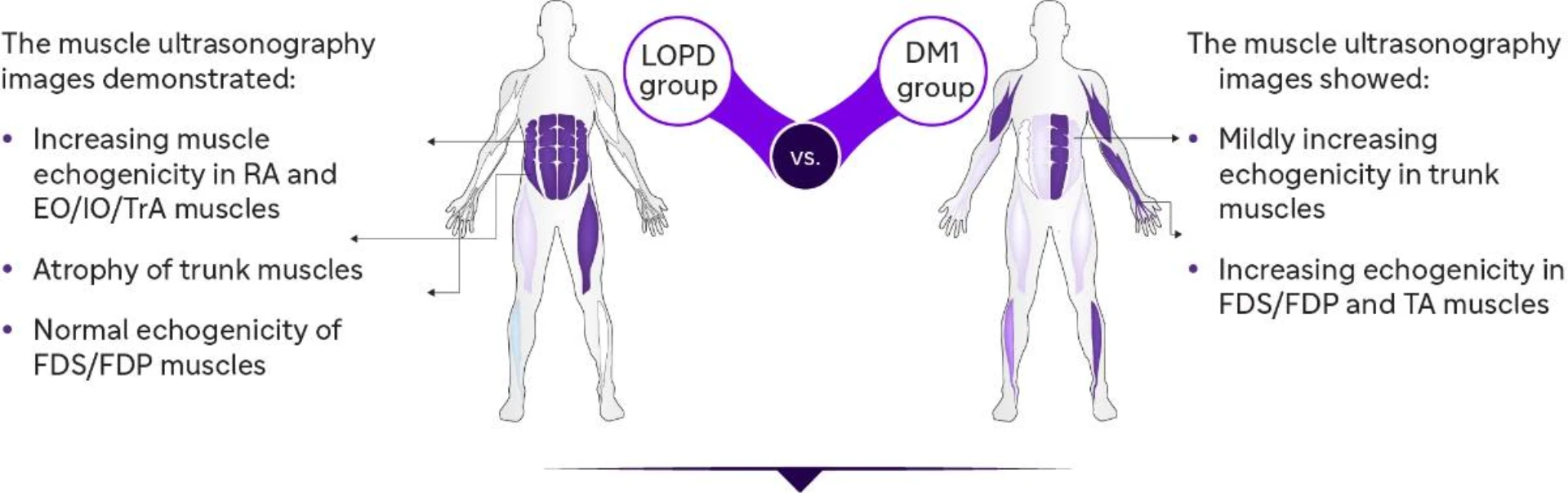
Qualitative echogenicity in skeletal muscles:
|
| Z Score | |
| Normal | <2 |
| Mild | 2-4 |
| Moderate | >4 |
| Severe | >6 |
|
Qualitative echogenicity in skeletal muscles:
|
Quantitative echogenicity and abdominal muscle thickness in muscular ultrasonography
Quantitative muscle echogenicity
.webp)
Trunk muscle thickness
.webp)
Total trunk grading sum score
%20(3).webp)
Conclusion
These findings suggest that muscle ultrasound is an efficient screening tool for:
- Assessing myopathic changes and disease-specific patterns
- Differential diagnosis of neuromuscular diseases
- Trunk muscles can be used for the differential diagnosis of LOPD and DM1.
- Muscle echography results correlated with clinical and motor functions.
- TIS can be used to investigate the trunk function
DM1: Myotonic dystrophy type 1; EO: External oblique; FDP: Flexor digitorum profundus; FDS: Flexor digitorum superficialis; IO: Internal oblique; LOPD: Late-onset Pompe disease; RA: Rectus abdominis; TA: Tibialis anterior; TIS: Trunk impairment scale; TrA: Transversus abdominis.
-
Hsieh PC, Chang CW, Ro LS, et al. Ultrasonography of abdominal muscles: Differential diagnosis of late-onset Pompe disease and myotonic dystrophy type 1. Front Neurol. 2022;13:944464.
MAT-KW-2300245 V1 Jul 2023