- Article
- Source: Campus Sanofi
- 14 May 2026
Atopic Dermatitis: Immune Endotypes and the OX40-OX40L Pathway
Introduction
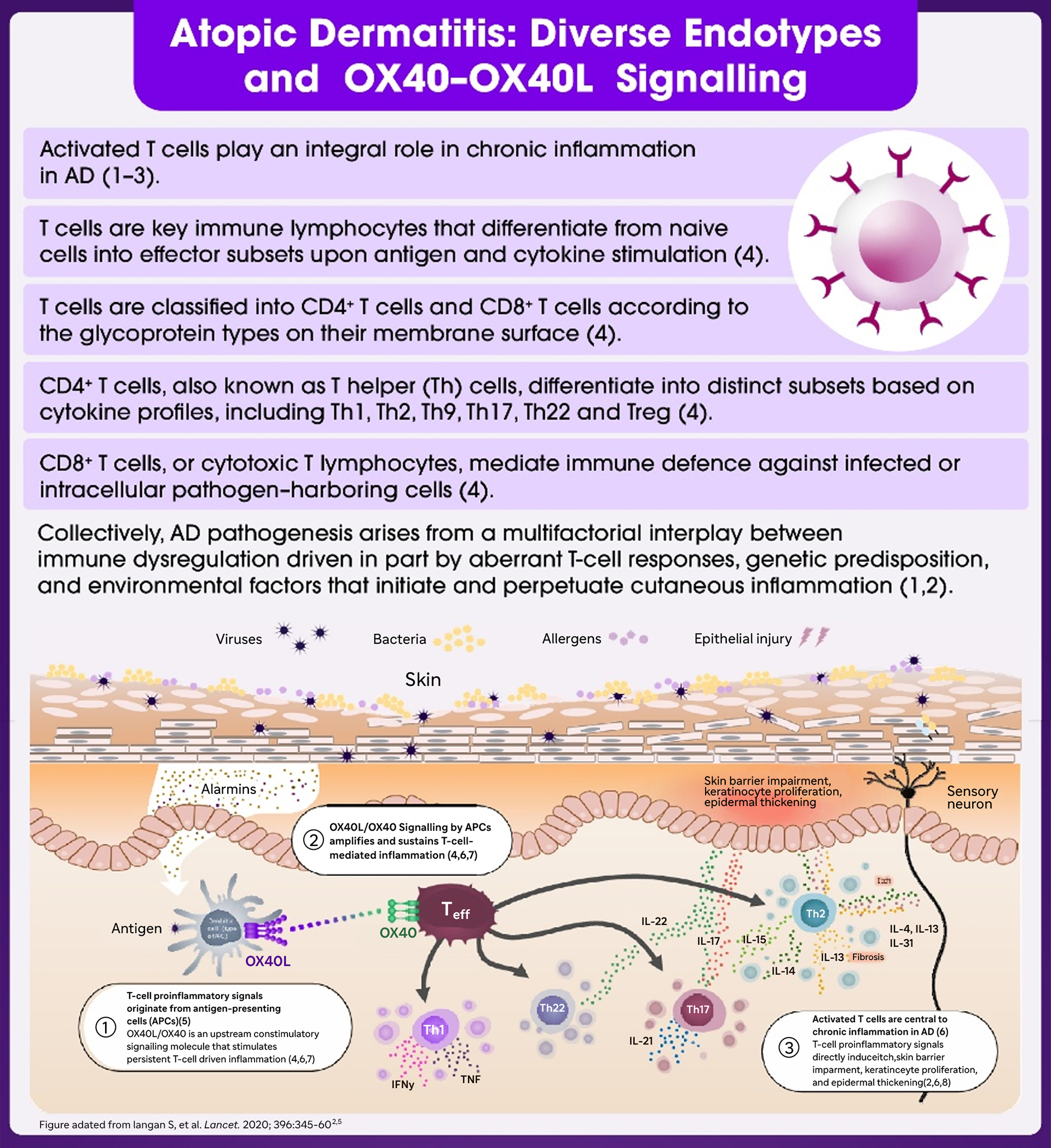
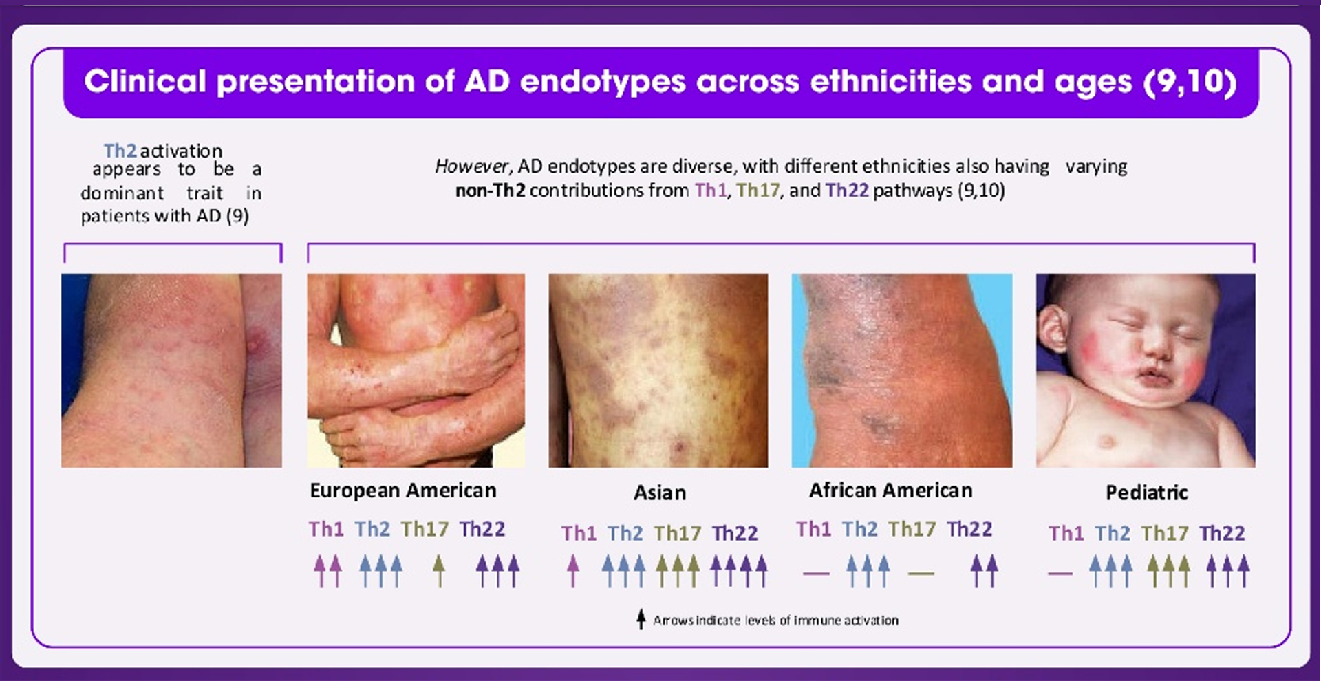
Atopic dermatitis (AD) is a common, chronic inflammatory skin disease that typically begins early in life (1). AD follows a relapsing–remitting course, and persistence of symptoms into adulthood contributes to its chronic nature, with up to 50% of affected children showing disease in later life (1). Globally, AD affects 20% of children and 10% of adults, with increasing incidence in industrialized countries (1). Loss of epithelial barrier integrity facilitates disease progression (1). During chronic inflammation, tumour necrosis factor receptor superfamily member 4; TNFRSF4 (OX40)-expressing T helper cells (Th1, Th17, and Th22) cells are recruited, and sustained OX40– OX40 ligand (OX40L) signalling promotes their proliferation (2). The resulting cytokines (IFN-γ, IL-17, IL-22) drive keratinocyte proliferation, epidermal thickening, and further immune cell recruitment, contributing to chronic AD (2).
Abbreviations:
AD – Atopic dermatitis; APC – Antigen-presenting cell; IgE – Immunoglobulin E; OX40 – Tumor necrosis factor receptor superfamily member 4 (CD134); OX40L – OX40 ligand (CD252); T cell – T lymphocyte; CD4⁺ – Cluster of differentiation 4 positive; Th – T helper cell; Th1 – Type 1 T helper cell; Th2 – Type 2 T helper cell; Th9 – Type 9 T helper cell; Th17 – Type 17 T helper cell; Th22 – Type 22 T helper cell; Treg – Regulatory T cell; IFN-γ – Interferon gamma; IL – Interleukin
- Kim MH, Kim SH, Park SY, Ban GY, Kim JH, Jung JW, et al. Characteristics of Adult Severe Refractory Asthma in Korea Analyzed From the Severe Asthma Registry. Allergy Asthma Immunol Res. 2019;11(1):43.
- Atopic dermatitis - The Lancet. The Lancet.
- Szalus K, Trzeciak M, Nowicki RJ. JAK-STAT Inhibitors in Atopic Dermatitis from Pathogenesis to Clinical Trials Results. Microorganisms. 2020 Nov 6;8(11):1743.
- Fu Y, Lin Q, Zhang Z, Zhang L. Therapeutic strategies for the costimulatory molecule OX40 in T-cell-mediated immunity. Acta Pharm Sin B. 2020 Mar;10(3):414–33.
- Tai Y, Wang Q, Korner H, Zhang L, Wei W. Molecular Mechanisms of T Cells Activation by Dendritic Cells in Autoimmune Diseases. Front Pharmacol. 2018 Jun 26;9:642.
- Furue M, Furue M. OX40L–OX40 Signaling in Atopic Dermatitis. J Clin Med. 2021 Jun 11;10(12):2578.
- Goronzy JJ, Weyand CM. T-cell co-stimulatory pathways in autoimmunity. Arthritis Res Ther. 2008;10(Suppl 1):S3.
- Kortekaas Krohn I, Aerts JL, Breckpot K, Govarts C, Knol E, Van Wijk F, et al. T-cell subsets in the skin and their role in inflammatory skin disorders. Allergy. 2022 Mar;77(3):827–42.
- Girolomoni G, De Bruin-Weller M, Aoki V, Kabashima K, Deleuran M, Puig L, et al. Nomenclature and clinical phenotypes of atopic dermatitis. Ther Adv Chronic Dis. 2021 Jan;12:20406223211002979.
- Czarnowicki T, He H, Krueger JG, Guttman-Yassky E. Atopic dermatitis endotypes and implications for targeted therapeutics. J Allergy Clin Immunol. 2019 Jan;143(1):1–11.
1. Mocanu M, Vâță D, Alexa AI, Trandafir L, Patrașcu AI, Hâncu MF, et al. Atopic Dermatitis—Beyond the Skin. Diagnostics. 2021 Aug 27;11(9):1553.
2. Sadrolashrafi K, Guo L, Kikuchi R, Hao A, Yamamoto RK, Tolson HC, et al. An OX-Tra’Ordinary Tale: The Role of OX40 and OX40L in Atopic Dermatitis. Cells. 2024 Mar 28;13(7):587.
MAT-SA-2600198-V1-APRIL2026