- Artikel
- Bron: Campus Sanofi
- 21 mei 2025
Diagnostic and confirmation testing

Enzyme assay
Gaucher disease is caused by a significant reduction in the enzyme acid β-glucosidase. Enzyme activity can be measured using a dried blood spot (DBS) sample, peripheral leukocytes, cultured fibroblasts, or amniotic fluid cells, with DBS being the preferred method in clinical practice. Enzyme assays do not distinguish between different types of Gaucher disease.
Molecular diagnostics
DNA tests can be used to aid in diagnosis as well as to identify carriers. Although many pathogenic variants associated with Gaucher disease have been identified, a negative result does not necessarily guarantee the absence of a Gaucher disease allele. However, there are some well-established associations that have been identified, particularly in the population of Ashkenazi Jews.2
Histology
The classic method for diagnosing Gaucher disease was the identification of Gaucher cells with lipid accumulation in bone marrow aspirate, liver or spleen biopsy, surgically collected. However, very similar cells (called pseudo-Gaucher cells) have been described to be associated with a number of other diseases, including:
- Chronic granulocytic leukemia
- Acute lymphocytic leukemia
- Lymphomas
- Thalassemia
- Multiple myeloma
- Hodgkin's Lymphoma3
Currently, biochemical and molecular techniques are more specific and less invasive, and routine histological evaluation is now discouraged.4
Biochemical markers3,4
Some macrophage-specific enzymes are elevated in the plasma of patients with Gaucher disease and some can be used as biomarkers in routine monitoring. Finding high levels of one or more of these markers is never enough to diagnose Gaucher disease, as levels of these markers can be high in other diseases. These biomarkers include:
- Glucosylsphingosine (Lyso-GL-1)
- Chitotriosidase
- Angiotensin-converting enzyme
- Tartrate-resistant acid phosphatase
In addition, high levels of ferritin, low vitamin B12, and hypocholesterolemia are typically found in people with Gaucher disease.
Lyso-GL-1 is recognized as a reliable and specific biomarker of Gaucher disease and is associated with the pathogenesis of the disease.5,6
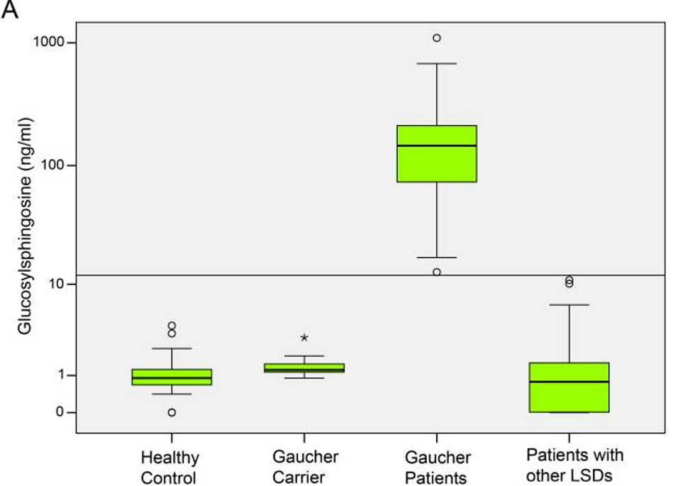
In the study of Rolfs A et al The Lyso-GL1 levels of 551 non-Jewish, Caucasian subjects were analysed. In total 148 healthy controls were compared to 98 genetically diagnosed GD patients, 13 GD carriers, and 262 patients with other LSDs (Niemann-Pick-Type C disease, Krabbe disease, Hunter disease and Fabry disease).5
In the figure the level of Lyso-GL1 is illustrated in the entire cohort. Lyso-GL1 in GD patients was compared to healthy controls, GD carriers and patients with other LSDs. The horizontal bar marks the cut-off for pathological Lyso-GL1 values above 12 ng/ml. Notably, only GD patients feature pathological values of Lyso-GL1.5
-
Machaczka M, Markuszewska-Kuczyńska A, Regenthal S, Jurczyszyn A, Gałązka K, Wahlin BE, Klimkowska M. Clinical utility of different bone marrow examination methods in the diagnosis of adults with sporadic Gaucher disease type 1. Pol Arch Med Wewn. 2014; 124(11):587-92.
-
Kaplan P, Baris H, De Meirleir L, Di Rocco M, El-Beshlawy A, Huemer M, et al. Revised recommendations for the management of Gaucher disease in children. Eur J Pediatr 2013;172:447-58.
-
Grabowski GA, Petsko GA, Kolodny EH. Gaucher Disease. In: Valle DL, Antonarakis S, Ballabio A, Beaudet AL, Mitchell GA. eds. The Online Metabolic and Molecular Bases of Inherited Disease. McGraw-Hill Education; 2019. Accessed October 06, 2025. https://ommbid.mhmedical.com/content.aspx?bookid=2709§ionid=225546056
-
Mistry PK, Cappellini M, Lukina E, et al. Consensus Conference: a reappraisal of Gaucher disease – diagnosis and disease management algorithms. Am J Hematol 2011; 86(1):110-115.
-
Rolfs A, et al. Glucosylsphingosine is a highly sensitive and specific biomarker for primary diagnostic and follow-up monitoring in gaucher disease in a non-Jewish, Caucasian cohort of Gaucher disease patients. PLoS One. 2013 Nov 20; 8(11):e79732.
-
Murugesan V, et al. Glucosylsphingosine is a key biomarker of Gaucher disease. Am j Hematol. 2016 Nov; 91(11):1082-89
Neem contact op

MAT-BE-2501473