- Article
- Source: Campus Sanofi
- Sep 15, 2026
Lung Disease in AATD: Potential Pulmonary Manifestations of Below-Normal AAT Levels
- Alpha-1 antitrypsin deficiency (AATD) is a genetic driven condition characterized by below-normal levels of the alpha-1 antitrypsin (AAT) protein1
- Individuals with below-normal AAT levels are predisposed to developing chronic obstructive pulmonary disease2
- Emphysema, a type of COPD, is a hallmark of AATD and the most common cause of mortality in patients with severe deficiency of the AAT protein1,3
- Other presentations of AATD include asthma with incompletely reversible airflow obstruction and uncontrolled bronchiectasis4
- As lung disease progresses in AATD, individuals face risk of decreased lung function and increased mortality1,5-7
- Risk of accelerated lung decline is exacerbated by factors such as a patient's environment (eg, pollutants and respiratory infections) and lifestyle (eg, smoking)8-10
The role of the alpha-1 antitrypsin (AAT) protein
The AAT protein plays a key role in helping protect lung tissue from irreversible damage by:
- Inhibiting a protease known as neutrophil elastase (NE)11
- Dampening pro-inflammatory cytokine production, inhibiting neutrophil activity11
- Reducing cellular damage from reactive oxygen species (ROS)11
In individuals living with AATD, pathogenic variants in the SERPINA1 gene can result in reduced circulating levels of AAT.12,13 Decreased levels of AAT may lead to uninhibited NE in the lungs, potentially resulting in emphysema.14-17 Exposure to smoke, air pollution, occupational inhalants, dust, and respiratory infections may further accelerate lung damage in people with below-normal AAT levels.8,10
AAT levels and pulmonary risk are closely linked
AAT protein levels are largely determined by genotype18
Range of plasma AAT levels based on allele combination19

Pathogenic variants in the SERPINA1 gene may lead to low or undetectable levels of serum AAT protein.12,13 Individuals with the MM genotype typically have normal levels of the AAT protein (20 μM to 53 μM), while those with the SZ or ZZ genotype often have substantially lower levels.19 More than 95% of all severely AAT-deficient individuals have either the ZZ or SZ genotype.5 Severe deficiency of the AAT protein is typically defined as ≤11 µM, which is the historic putative threshold.6
The risk for lung disease generally increases as AAT levels decrease. However, risk varies and may be influenced by genotype, smoking history, environmental and occupational exposures, and pathophysiologic factors.8,10,19 Even heterozygous genotypes, such as MZ and SZ, may be associated with increased pulmonary risk.18,19
aAAT levels may also be measured as mg/dL. MM: 90-200 mg/dL; MZ: 66-120 mg/dL; SZ: 45-80 mg/dL; ZZ: 10-40 mg/dL.19,20
bThis is not a comprehensive list of genotypes. Patients may also be affected by additional genotypes, including F, I, and Q0/null.
AATD is a genetic contributor of progressive lung disease, such as COPD5,10,21
AATD is the best-known genetic risk factor for COPD and requires disease-specific management.5,21 Yet, due to the overlapping clinical presentations, AATD is often incompletely diagnosed as COPD.22-24
It is estimated that 1% of patients with COPD have severe AAT deficiency.17 However, >90% of individuals with AATD remain undiagnosed.25
Because symptoms overlap significantly, clinical presentation alone is not enough to distinguish COPD caused by AATD from non-genetic COPD.26 Overlapping symptoms include1:
- Shortness of breath during daily activities
- Wheezing
- Decreased ability to exercise
- Excessive mucus production while coughing
Without testing, patients with AATD may remain unidentified and at continued risk for disease progression.27,28 Guidelines recommend testing for AATD in all patients with1,5:
- COPD
- Emphysema
- Asthma with incompletely reversible airflow obstruction
- Unexplained bronchiectasis
An imbalance between protease(s) and AAT activity allows ongoing protease-mediated injury, often subclinical, to accumulate over time and contribute to the development of emphysema.15-17
AATD and emphysema
The lower AAT levels are, the higher the risk of AATD-related emphysema19
Emphysema is the hallmark pulmonary manifestation of AATD.17,20 AATD-associated emphysema is typically panacinar (panlobular), which typically affects the lower lung zones more prominently than smoking-related emphysema (centrilobular emphysema).17,20 Emphysema is the most common cause of mortality in patients with severe deficiency of AAT, contributing to up to 72% of deaths.3,a
Emphysema is reported in 47% of Medicare patients with AATD-related COPD29,b

aOf the 1129 Registry enrollees, 204 subjects (18.1%) died over the course of follow-up. Of the 204 deaths, records were available for 120 subjects. Underlying cause of death was ascertained in 118 of the 120 subjects with records. In the overall cohort of 204 decedents, 13.7% were never-smokers, 86.3% were ever-smokers.
3
bRetrospective analysis of 742 Medicare patients with AATD with emphysema and COPD exacerbations compared with 7420 matched individuals with COPD without AATD (2015-2021).29
Beyond COPD and emphysema: Other pulmonary manifestations
AATD can present in multiple ways
In addition to COPD and emphysema, patients with AATD may experience1,17:
- Asthma
- Bronchiectasis
- Chronic cough
- Wheezing
- Excess phlegm production
- Bronchial hyperresponsiveness
AATD is linked to impaired lung function and increasing mortality
Patients with AATD may be at increased risk of irreversible lung function decline due to emphysema progression.5,6
In a study of 74 patients with AATD (ZZ, n=33; SZ, n=41)7:
- Patients with ZZ showed greater annual lung functional decline (FEV1/DLCO/KCO) and densitometric parameters (PD-15/HU-950)
- Smoking and exacerbations were associated with a greater functional decline (FEV1/DLCO/KCO) and densitometric parameters
- Significant lung-basal region loss occurred, with greater decline in advanced stages
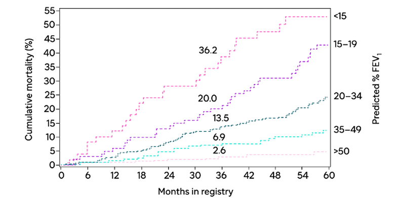
Lower predicted FEV1 is strongly associated with higher cumulative mortality over time3
Kaplan-Meier mortality analysis by predicted % FEV13
Routinely monitor patients with AATD and consider ongoing assessment in individuals with genotypes associated with below-normal AATD.
With AATD, pulmonary risks are multifactorial and vary among individuals
Early diagnosis and regular monitoring may help improve the prognosis of AATD
The AAT protein plays a critical role in protecting lung tissue from protease-mediated damage.13 When pathogenic SERPINA1 variants result in below-normal AAT levels, the lungs may become increasingly vulnerable to progressive injury.12,13 The pulmonary impact of AATD varies among individuals and is influenced by genotype, circulating AAT protein levels, smoking status, environmental and occupational exposures, and disease history.18 Over time, AATD may contribute to emphysema, COPD, and other pulmonary manifestations that can increase overall disease burden.8
Interested in learning more?
FAQs
While both AATD and cystic fibrosis are genetic driven disorders that affect pulmonary health, they have distinct pathologies. AATD is caused by pathogenic mutations in the SERPINA1 gene that can lead to decreased levels of the AAT protein, with chronic obstructive pulmonary symptoms that typically present in adulthood. Cystic fibrosis is caused by mutations to the CFTR gene, which regulates the cellular exchange of water and salt, leading to sticky, thick mucus buildup in the lungs, pancreas, and other organs.3,12,13,30
Pulmonary risks in AATD are influenced by multiple factors beyond smoking exposure. Genotype, circulating AAT protein levels, and environmental and occupational exposures all contribute to lung disease risk. While smoking is a major risk factor for accelerated lung decline, patients with below-normal AAT levels may be more vulnerable to protease-mediated lung damage, resulting in different clinical outcomes despite similar smoking histories.18,31
Normal serum AAT levels associated with the MM genotype range from 20 μM to 53 μM. As AAT levels decrease, the lungs may become increasingly susceptible to protease-mediated tissue damage. Severe deficiency of AAT is considered to be ≤11 µM, which is also the historic putative threshold. Because pulmonary risk exists along a continuum and varies between individuals, AAT levels should be interpreted alongside genotype, clinical history, and other risk factors.6,19
AATD is often underrecognized because its pulmonary manifestations overlap substantially with those of COPD. Symptoms such as shortness of breath, wheezing, chronic cough, mucus production, and reduced exercise tolerance are common to both conditions. As a result, some patients receive a diagnosis of COPD without evaluation for an underlying genetic cause. Since clinical presentation alone cannot distinguish AATD-related COPD from non-genetic COPD, guidelines (ATS and GOLD) recommend parallel genetic and serum level testing in all patients with COPD for AATD.1,22-24,32
Yes. Although smoking can accelerate lung damage in individuals with AATD, pulmonary disease can develop even in patients with little or no smoking exposure. Reduced AAT levels may leave the lungs vulnerable to neutrophil elastase-mediated tissue injury, increasing the risk of emphysema and other pulmonary manifestations. Environmental exposures, such as air pollution or occupational dust, may also contribute to pulmonary risks.8
Genotype is an important determinant of AAT protein levels and pulmonary risk, but it does not fully predict disease severity. Individuals with genotypes associated with below-normal AAT levels, such as ZZ and SZ, generally face a higher risk of lung disease and progression. However, pulmonary outcomes are also influenced by smoking history, environmental and occupational exposures, exacerbations, and other patient-specific factors.1,7,18,19
Yes. Patients with the same genotype may experience markedly different pulmonary outcomes. Although genotype strongly influences AAT levels and disease risk, other factors, including smoking status, environmental and occupational exposures, respiratory infections, exacerbation frequency, and baseline lung health can affect the trajectory of disease. As a result, patients with the same genotype may differ in symptom burden, rate of lung function decline, and overall disease severity.8,18,31
Definitions: ATS=American Thoracic Society; COPD=chronic obstructive pulmonary disease; DLCO=diffusing capacity of the lungs for carbon monoxide; FEV1=forced expiratory volume in 1 second; GOLD=Global Initiative for Chronic Obstructive Lung Disease; HU-950=Hounsfield units less than 950; KCO=carbon monoxide transfer coefficient; PD-15=15 percentile lung density.
References: 1. American Thoracic Society; European Respiratory Society. American Thoracic Society/European Respiratory Society statement: standards for the diagnosis and management of individuals with alpha-1 antitrypsin deficiency. Am J Respir Crit Care Med. 2003;168(7):818-900. 2. Teckman JH, Blomenkamp KS. Pathophysiology of alpha-1 antitrypsin deficiency liver disease. In: Borel F, Mueller C, eds. Alpha-1 Antitrypsin Deficiency: Methods and Protocols. Humana Press; 2017:1-8. 3. Stoller JK, Tomashefski J Jr, Crystal RG, et al. Mortality in individuals with severe deficiency of α1-antitrypsin: findings from the National Heart, Lung, and Blood Institute Registry. Chest. 2005;127(4):1196-1204. 4. Craig TJ, Corbett ML, Meadows JA. Improving detection of alpha-1 antitrypsin deficiency: role of the allergist. J Allergy Clin Immunol Pract. 2023;11(8):2348-2354. 5. Sandhaus RA, Turino G, Brantly ML, et al. The diagnosis and management of alpha-1 antitrypsin deficiency in the adult. Chronic Obstr Pulm Dis. 2016;3(3):668-682. 6. Barjaktarevic I, Campos M. Management of lung disease in alpha-1 antitrypsin deficiency: what we do and what we do not know. Ther Adv Chronic Dis. 2021;12:49-63. 7. Esmaili S, Rodriguez Hermosa JL, Centanaro GV, Alvarez-Sala JL, Esmaili I, Rubio MC. Progression and augmentation therapy in PiSZ and PiZZ alpha-1 antitrypsin deficiency: a longitudinal functional and densitometric study. Biomolecules. 2025;15(4):599. doi:10.3390/biom15040599 8. Torres-Durán M, Lopez-Campos JL, Barrecheguren M, et al. Alpha-1 antitrypsin deficiency: outstanding questions and future directions. Orphanet J Rare Dis. 2018;13(1):114. doi:10.1186/s13023-018-0856-9 9. Wang T, Shuai P, Wang Q, et al. α‑1 Antitrypsin is a potential target of inflammation and immunomodulation (Review). Mol Med Rep. 2025;31(4):107. doi:10.3892/mmr.2025.13472 10. Kokturk N, Khodayari N, Lascano J, et al. Lung inflammation in alpha‑1‑antitrypsin deficient individuals with normal lung function. Respir Res. 2023;24(1):40. doi:10.1186/s12931-023-02343-3 11. Mazzuca C, Vitiello L, Travaglini S, et al. Immunological and homeostatic pathways of alpha-1 antitrypsin: a new therapeutic potential. Front Immunol. 2024;15:1443297. doi:10.3389/fimmu.2024.1443297 12. Cazzola M, Stolz D, Rogliani P, Matera MG. α1-Antitrypsin deficiency and chronic respiratory disorders. Eur Respir Rev. 2020;29(155):190073. doi:10.1183/16000617.0073-2019 13. Karatas E, Bouchecareilh M. Alpha 1-antitrypsin deficiency: a disorder of proteostasis-mediated protein folding and trafficking pathways. Int J Mol Sci. 2020;21(4):1493. doi:10.3390/ijms21041493 14. University of Utah Health. Alpha-1 antitrypsin deficiency. Accessed February 6, 2026. https://learn.genetics.utah.edu/content/genetics/alpha1/ 15. Viglio S, Iadarola P, D’Amato M, Stolk J. Methods of purification and application procedures of alpha1 antitrypsin: a long-lasting history. Molecules. 2020;25(17):4014. 16. Abdulkarim A, Craig TJ. Alpha-1 antitrypsin mutation (archived). In: StatPearls [Internet]. StatPearls Publishing; September 26, 2022. Accessed February 20, 2026. https://www.ncbi.nlm.nih.gov/books/NBK482180 17. Kalfopoulos M, Wetmore K, ElMallah MK. Pathophysiology of alpha-1 antitrypsin lung disease. In: Borel F, Mueller C, eds. Alpha-1 Antitrypsin Deficiency: Methods in Molecular Biology. Humana Press; 2017:9-20. 18. Feitosa PHR, de Oliveira Castellano MVC, da Costa CH, et al. Recommendations for the diagnosis and treatment of alpha-1 antitrypsin deficiency. J Bras Pneumol. 2024;50(5):e20240235. doi:10.36416/1806-3756/e20240235 19. Mulkareddy V, Roman J. Pulmonary manifestations of alpha 1 antitrypsin deficiency. Am J Med Sci. 2024;368(1):1-8. 20. Strnad P, McElvaney NG, Lomas DA. Alpha1-antitrypsin deficiency. N Engl J Med. 2020;382(15):1443-1455. 21. COPD Foundation. Genetic COPD. Reviewed April 17, 2025. Accessed July 29, 2026. https://www.copdfoundation.org/Learn-More/I-am-a-Healthcare-Provider/Genetic-COPD.aspx 22. Quinn M, Ellis P, Pye A, Turner AM. Obstacles to early diagnosis and treatment of alpha-1 antitrypsin deficiency: current perspectives. Ther Clin Risk Manag. 2020;16:1243-1255. 23. Global Initiative for Chronic Obstructive Lung Disease (GOLD). GOLD Report. Accessed February 3, 2026. https://goldcopd.org/2026-gold-report-and-pocket-guide/ 24. Campos MA, Wanner A, Zhang G, Sandhaus RA. Trends in the diagnosis of symptomatic patients with alpha1-antitrypsin deficiency between 1968 and 2003. Chest. 2005;128(3):1179-1186. 25. Craig TJ, Henao MP. Advances in managing COPD related to α1 – antitrypsin deficiency: an under-recognized genetic disorder. Allergy. 2018;73(11):2110-2121. 26. Demeo DL, Sandhaus RA, Barker AF, et al. Determinants of airflow obstruction in severe alpha-1-antitrypsin deficiency. Thorax. 2007;62(9):806-813. 27. Tejwani V, Nowacki AS, Fye E, Sanders C, Stoller JK. The impact of delayed diagnosis of alpha-1 antitrypsin deficiency: the association between diagnostic delay and worsened clinical status. Respir Care. 2019;64(8):915-922. 28. Meischl T, Schmid-Scherzer K, Vafai-Tabrizi F, et al. The impact of diagnostic delay on survival in alpha-1-antitrypsin deficiency: results from the Austrian alpha-1 lung registry. Respir Res. 2023;24(1):34. doi:10.1186/s12931-023-02338-0 29. Khandelwal N, Hinson J,Nguyen T, et al. Clinical and economic outcomes in patients with alpha-1 antitrypsin deficiency in a US Medicare advantage population. J Health Econ Outcomes Res. 2025;12(1):66-74. 30. Ratjen F, Bell SC, Rowe SM, Goss CH, Quittner AL, Bush A. Cystic fibrosis. Nat Rev Dis Primers. 2015;1:15010. doi:10.1038/nrdp.2015.10 31. Molloy K, Hersh CP, Morris VB, et al. Clarification of the risk of chronic obstructive pulmonary disease in α1-antitrypsin deficiency PiMZ heterozygotes. Am J Respir Crit Care Med. 2014;189(4):419-427. 32. McElvaney GN, Sandhaus RA, Miravitlles M, et al. Clinical considerations in individuals with α1-antitrypsin PI*SZ genotype. Eur Respir J. 2020;55(6):1902410 doi:10.1183/13993003.02410-2019
MAT-US-2609068-v1.0-09/2026. Last Updated: September 2026
